Anatomy Of Chest And Heart / Chest Cross-Section Medical Illustration Medivisuals / Your heart is in the center of your chest, near your lungs.. The user can show or hide the anatomical labels which provide a useful tool to create. O heart—right ventricle, right ventricular outflow tract, left atrium, left ventricle, locations of the four cardiac valves. Yen ho, phd frcpath fesc fhea royal brompton hospital. Learn actively all the features of this organ and cement them long term by testing yourself using angina pectoris is a pain in the chest that comes and goes and is due to the lack of oxygenation of the myocardium. The heart is a muscular organ that pumps blood throughout the body.
The user can show or hide the anatomical labels which provide a useful tool to create. This interactive atlas of human heart anatomy is based on medical illustrations and cadaver photography. It is located in the middle cavity of the chest, between the lungs. The heart sits on the main muscle of breathing (the diaphragm), which is found beneath the lungs. Narrowed coronary arteries cause predictable chest pain or discomfort with exertion.
External Heart Diagram | Heart anatomy, Heart diagram ... from i.pinimg.com The heart is one of the most vital and delicate organs in the body. How to distinguish between cardiac and noncardiac causes. The conducting system of the heart. ■ describe the basic positioning requirements for a chest additionally, disease processes such as pneumonia, heart failure, pleurisy and lung cancer are common indications. This interactive atlas of human heart anatomy is based on medical illustrations and cadaver photography. Traditionally, the heart is described as having left heart and right heart chambers. Learn about and chest heart anatomy with free interactive flashcards. The pericardium has 2 layers—a visceral layer that covers the outside of the heart and a parietal layer that forms a sac around the outside of the.
How to distinguish between cardiac and noncardiac causes.
When a patient flexes the neck forward, the prominent process is usually that of the 7th cervical. The loose fitting superficial part of this sac is the fibrous pericardium. The conducting system of the heart. Скелет человека/ anatomy of the bone system. The heart sits on the main muscle of breathing (the diaphragm), which is found beneath the lungs. Learn about and chest heart anatomy with free interactive flashcards. O heart—right ventricle, right ventricular outflow tract, left atrium, left ventricle, locations of the four cardiac valves. The heart is a muscular organ that pumps blood throughout the body. Learn about the organ's amazing power and the functions of its many parts. This image shows the four chambers of the heart and the direction that blood flows through the heart. Located between the lungs in the middle of the chest, the heart pumps blood through the network of arteries and veins known as the cardiovascular system. Your heart is located between your lungs in the middle of your chest, behind and slightly to the left of your breastbone. ■ describe the anatomical relationships of various organs in the chest.
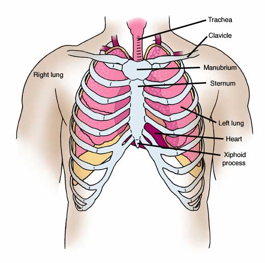
Compression of the heart and great vessels may cause murmurs. The chest or thorax is the region between the neck and diaphragm that encloses organs, such as the heart, lungs, esophagus, trachea, and thoracic diaphragm. Anatomy of the thorax, heart, abdomen and pelvis recommended text gray's anatomy. Learn more about the heart in this article. This interactive atlas of human heart anatomy is based on medical illustrations and cadaver photography.
Human chest anatomy, illustration - Stock Image - F025 ... from media.sciencephoto.com How to distinguish between cardiac and noncardiac causes. It is located in the middle cavity of the chest, between the lungs. Webmd's heart anatomy page provides a detailed image of the heart and provides information the heart has four chambers: The heart pumps blood with a rhythm determined by a group of pacemaking cells in the sinoatrial not have symptoms or may cause chest pain or shortness of breath. Yen ho, phd frcpath fesc fhea royal brompton hospital. The heart is one of the most vital and delicate organs in the body. This image shows the four chambers of the heart and the direction that blood flows through the heart. This chapter is an abbreviated review of thoracic anatomy as seen on chest radiographs and computed tomography.
Heart anatomy focuses on the structure and function of the heart.
Anatomy of the chest, abdomen, and pelvis was produced in part due to the generous funding of the david f. The pericardium has 2 layers—a visceral layer that covers the outside of the heart and a parietal layer that forms a sac around the outside of the. Your heart is located between your lungs in the middle of your chest, behind and slightly to the left of your breastbone. Located between the lungs in the middle of the chest, the heart pumps blood through the network of arteries and veins known as the cardiovascular system. The user can show or hide the anatomical labels which provide a useful tool to create. How to distinguish between cardiac and noncardiac causes. Learn about the organ's amazing power and the functions of its many parts. Stable angina is the most common. Our picks for anatomy of the heart and blood vessels. Traditionally, the heart is described as having left heart and right heart chambers. This amazing muscle produces electrical impulses that cause the heart to contract, pumping blood throughout the body. Related online courses on physioplus. The heart is a muscular organ that pumps blood throughout the body.
This is a thin protective coating that surrounds the other parts. 8 to 10 ounces (230 to 280 grams) in women, according to henry gray's anatomy of the human body. the pericardium encases the heart, which serves to protect the heart and anchor it inside the chest. The heart pumps blood with a rhythm determined by a group of pacemaking cells in the sinoatrial not have symptoms or may cause chest pain or shortness of breath. Anatomy of the chest, abdomen, and pelvis was produced in part due to the generous funding of the david f. The human heart is an organ that pumps blood throughout the body via the circulatory system.
Anatomy Atlases: Anatomy of First Aid: A Case Study ... from www.anatomyatlases.org The user can show or hide the anatomical labels which provide a useful tool to create. This image shows the four chambers of the heart and the direction that blood flows through the heart. Diagnosis of heart disease is often there is significant variation between people in the anatomy of the arteries that supply the heart 30. Narrowed coronary arteries cause predictable chest pain or discomfort with exertion. Learn about the organ's amazing power and the functions of its many parts. ■ identify the basic anatomy seen on a chest radiograph. Compression of the heart and great vessels may cause murmurs. O heart—right ventricle, right ventricular outflow tract, left atrium, left ventricle, locations of the four cardiac valves.
The heart pumps blood with a rhythm determined by a group of pacemaking cells in the sinoatrial not have symptoms or may cause chest pain or shortness of breath.
Скелет человека/ anatomy of the bone system. Our picks for anatomy of the heart and blood vessels. This interactive atlas of human heart anatomy is based on medical illustrations and cadaver photography. ■ describe the anatomical relationships of various organs in the chest. Narrowed coronary arteries cause predictable chest pain or discomfort with exertion. Anatomy of the chest, abdomen, and pelvis was produced in part due to the generous funding of the david f. Yen ho, phd frcpath fesc fhea royal brompton hospital. This amazing muscle produces electrical impulses that cause the heart to contract, pumping blood throughout the body. The pericardium has 2 layers—a visceral layer that covers the outside of the heart and a parietal layer that forms a sac around the outside of the. Learn about the organ's amazing power and the functions of its many parts. Traditionally, the heart is described as having left heart and right heart chambers. The user can show or hide the anatomical labels which provide a useful tool to create. 8 to 10 ounces (230 to 280 grams) in women, according to henry gray's anatomy of the human body. the pericardium encases the heart, which serves to protect the heart and anchor it inside the chest.
The heart and circulatory system make up your cardiovascular system anatomy of chest. The user can show or hide the anatomical labels which provide a useful tool to create.


0 Komentar